
EKG's
A primer for patients
What is an EKG and Why Your Doctor Orders It?
An Electrocardiogram (EKG or ECG) is a simple, painless test that measures the electrical activity of your heart. It's like taking a snapshot of your heart's rhythm and can provide valuable information about your heart's health.
How Does an EKG Work?
During an EKG, small electrodes are attached to your chest, arms, and legs. These electrodes detect the electrical signals generated by your heart as it beats. The signals are then recorded as waves on a paper or displayed on a monitor. Multiple wires are used so the doctor can get a view of the electrical pattern fro multiple angles
What Does an EKG Measure?
An EKG measures several aspects of your heart's electrical activity, including:
Why Does Your Doctor Order an EKG?
Your doctor may order an EKG for various reasons:
An EKG is a valuable tool that helps your doctor assess your heart's electrical activity, detect abnormalities, and monitor your heart's health over time. It's a quick and non-invasive test that can provide important information to guide your healthcare. If your doctor recommends an EKG, don't hesitate to ask questions and discuss any concerns you may have. Your heart health is important, and an EKG is one way to ensure it stays in rhythm.
How Does an EKG Work?
During an EKG, small electrodes are attached to your chest, arms, and legs. These electrodes detect the electrical signals generated by your heart as it beats. The signals are then recorded as waves on a paper or displayed on a monitor. Multiple wires are used so the doctor can get a view of the electrical pattern fro multiple angles
What Does an EKG Measure?
An EKG measures several aspects of your heart's electrical activity, including:
- Heart Rate: It shows how fast or slow your heart is beating.
- Rhythm: It reveals whether your heart rhythm is regular or irregular. This helps detect abnormalities like atrial fibrillation or abnormal heart rhythms.
- Conduction System: It assesses the pathways that electrical impulses follow through your heart, detecting any blocks or abnormalities.
- Heart Muscle Health: Certain patterns on the EKG can indicate if parts of your heart are damaged or not receiving enough oxygen.
Why Does Your Doctor Order an EKG?
Your doctor may order an EKG for various reasons:
- Routine Check-Up: As part of a routine physical exam, especially if you have risk factors for heart disease, like high blood pressure, diabetes, or a family history of heart problems.
- Symptoms: If you experience symptoms like chest pain, shortness of breath, palpitations (feeling of rapid or irregular heartbeat), dizziness, or fainting, an EKG helps identify the cause.
- Monitoring Treatment: To monitor the effectiveness of medications or treatments for heart conditions.
- Baseline Assessment: Before undergoing surgery or starting certain medications, your doctor may request an EKG to establish a baseline of your heart's health.
An EKG is a valuable tool that helps your doctor assess your heart's electrical activity, detect abnormalities, and monitor your heart's health over time. It's a quick and non-invasive test that can provide important information to guide your healthcare. If your doctor recommends an EKG, don't hesitate to ask questions and discuss any concerns you may have. Your heart health is important, and an EKG is one way to ensure it stays in rhythm.
So what do all those lines and bumps on my EKG mean
Introduction:
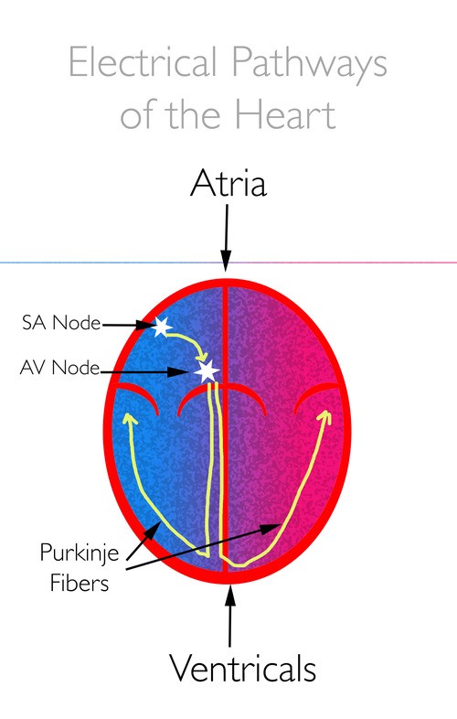
Your heart's steady beat is like a well-orchestrated symphony conducted by its own electrical system. Understanding how this electrical impulse travels through your heart, from the SA node to the rest of the heart, can provide valuable insights into your cardiovascular health. Let's explore this fascinating journey and how it's reflected on an electrocardiogram (EKG) tracing.
The SA Node: Initiating the Beat
Imagine the sinoatrial (SA) node as the conductor of your heart's orchestra. It's the heart's natural pacemaker. Located in the right atrium, it generates electrical impulses, signaling the heart to beat. These signals spread like ripples in a pond, causing the atria to contract and push blood into the ventricles.
The AV Node: Regulating the Pace
The electrical impulses then travel to the atrioventricular (AV) node, situated between the atria and ventricles. Think of it as a traffic cop regulating the flow of signals. The AV node briefly delays the impulse, allowing the ventricles time to fill with blood before contracting. This delay ensures efficient blood flow and coordination between the upper and lower chambers of the heart.
Propagation Through the Heart's Pathways:
After passing through the AV node, the electrical impulse races along specialized pathways known as the bundle of His and Purkinje fibers. These pathways act like high-speed highways, rapidly transmitting the signal to the ventricles. As a result, the ventricles contract, pumping blood to the lungs and the rest of the body.
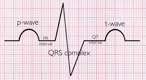
Correlation with EKG Tracing:
An electrocardiogram (EKG or ECG) is a graphic representation of your heart's electrical activity. Understanding how the electrical impulse travels through your heart helps interpret the various waves and intervals observed on an EKG tracing:
EKG Terminology
Limitations of the EKG
While an electrocardiogram (EKG or ECG) is a valuable tool for detecting and diagnosing various heart conditions, it does have limitations, especially when used in isolation or without proper interpretation by a trained healthcare professional. Here are some limitations of an EKG for an average person:
Despite these limitations, an EKG remains a valuable diagnostic tool when used in conjunction with clinical evaluation, patient history, and other diagnostic tests to assess cardiac health and guide treatment decisions.
Conclusion:
Your heart's electrical system orchestrates each beat, ensuring the efficient pumping of blood throughout your body. By understanding this process and its representation on an EKG tracing, we gain insight into your heart's health and function. If you have any questions or concerns about your heart's electrical activity or EKG results, don't hesitate to discuss them with your healthcare provider. Your heart's rhythm is the melody of your life, and it's essential to keep it in harmony.
An electrocardiogram (EKG or ECG) is a graphic representation of your heart's electrical activity. Understanding how the electrical impulse travels through your heart helps interpret the various waves and intervals observed on an EKG tracing:
- P Wave: The P wave corresponds to atrial depolarization, initiated by the SA node. It represents the electrical activity as the atria contract, pushing blood into the ventricles.
- PR Interval: The PR interval reflects the time it takes for the electrical impulse to travel from the SA node to the AV node. It includes the delay at the AV node, ensuring sequential contraction of the atria and ventricles.
- QRS Complex: The QRS complex signifies ventricular depolarization, triggered by the electrical signal transmitted through the bundle of His and Purkinje fibers. It corresponds to the contraction of the ventricles, leading to ejection of blood into the arteries.
- T Wave: The T wave represents ventricular repolarization, indicating the resetting of the electrical charge of the ventricles after contraction. we can't see the wave produced when the atria repolarize because its small and buried inside the QRS complex
EKG Terminology
- Bradycardia - A heart rate that is slower than the normal 60-100 beats per minute. This can be normal for many people especially if they exercise a lot or if they are on certain medications like beta blockers
- Tachycardia - A heart rate that is faster than the normal 60-100 beats per minute. This can happen in response to stress, anxiety, or exertion but can also be seen in conditions like dehydration, hyperthyroidism, and when there has been a great loss of blood.
- Sinus Rhythm - This means that the beat of the heart is being triggered by the SA node, the hearts natural pacemaker. This is the normal rhythm of a heart
- Sinus Arrhythmia - Again, a rhythm dictated and sent out by the SA node but with slightly irregular intervals. This is not harmful and may be completely normal for some people.
- VPC (Ventricular Premature Contraction) i- This s a type of irregular heartbeat originating in the heart's lower chambers. It feels like a sudden, extra heartbeat or flutter. Sporadic VPC's are generally harmless,
- APC (Atrial Premature Contraction) - This is an early heartbeat originating in the heart's upper chambers. It may feel like a "skipped" beat or flutter. This is nearly always benign,
- Atrial Fibrillation - This is the most common type of irregular heart rhythm. In atrial fibrillation, the electrical signals in the atria become disorganized and rapid, causing the atria to quiver or fibrillate instead of contracting effectively. This chaotic electrical activity results in irregular and often rapid heartbeats. The irregular electrical impulses bombard the AV node, which tries to regulate the impulses it sends to the ventricles. However, due to the irregularity and rapidity of the signals from the atria, the ventricles also beat irregularly and sometimes rapidly. Because the atria don't contract effectively blood can pool in areas and stagnate leading to clots which can break off and cause a stroke.
- ST-T wave changes - These are deviations in the shape or position of the T-wave or the elevation or depression of the QT interval line. These can take many forms. Most are non-specific and have no health implications while some can indicate a possible heart attack. Doctors can often clarify borderline cases by comparing to prior EKG's the patient may have had.
Limitations of the EKG
While an electrocardiogram (EKG or ECG) is a valuable tool for detecting and diagnosing various heart conditions, it does have limitations, especially when used in isolation or without proper interpretation by a trained healthcare professional. Here are some limitations of an EKG for an average person:
- Intermittent Detection: An EKG records the heart's electrical activity only at the time of the test. It may not capture intermittent or transient abnormalities that occur between tests. Some heart conditions, such as paroxysmal atrial fibrillation which comes and goes during irregular intervals, may not be detected if the episode does not occur during the EKG recording. In some cases, specialized monitoring techniques such as Holter monitoring, event recorders, or implantable loop recorders may be necessary for accurate diagnosis.
- False Positives and Negatives: An EKG may produce false-positive or false-negative results. False positives occur when the EKG suggests the presence of a heart condition that is not actually present. False negatives occur when the EKG fails to detect a heart condition that is present. Factors such as poor electrode placement, patient movement during the test, or technical errors can contribute to inaccurate results.
- Non-specific Findings: Some EKG findings may be non-specific, meaning they can be indicative of various underlying conditions. For example, ST-segment and T-wave changes can occur due to myocardial ischemia, electrolyte imbalances, or other factors, and they can be completely normal for some people making it challenging to pinpoint the exact cause without an old EKG for comparison or additional testing or clinical evaluation.
- Influence of Other Factors: Certain factors such as medication use, electrolyte imbalances, underlying medical conditions, and patient characteristics (e.g., obesity, lung disease) can affect EKG results. Healthcare providers need to consider these factors when interpreting EKG findings.
Despite these limitations, an EKG remains a valuable diagnostic tool when used in conjunction with clinical evaluation, patient history, and other diagnostic tests to assess cardiac health and guide treatment decisions.
Conclusion:
Your heart's electrical system orchestrates each beat, ensuring the efficient pumping of blood throughout your body. By understanding this process and its representation on an EKG tracing, we gain insight into your heart's health and function. If you have any questions or concerns about your heart's electrical activity or EKG results, don't hesitate to discuss them with your healthcare provider. Your heart's rhythm is the melody of your life, and it's essential to keep it in harmony.